The Plasma Membrane
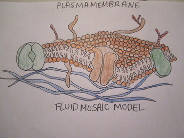
-The plasma membrane is represented by a model referred to as the fluid mosaic model.
-It has three different names:
*the plasma membrane
*cell membrane
*phospholipid bilayer
-On the membrane there are phospholipids, protein carriers, ion channels, glycolipids, and glycoproteins.
-Phospholipids make up the membrane. They have a head, and two tails. These are double layered, tails facing inwards. The head is hydrophilic and the tails are hydrophobic.
-Protein carriers: change their shape to allow through large or charged molecules because these cannot get through the phospholipids.
-Ion channels: Allow large or charged molecules through the channel in the middle of them.
-Glycolipids and glycoproteins: are both receptors for the cell. They communicate with other cells. The only difference is that a glycolipid is connected to a fat (lipid), and a glycoprotein is attached to a protein.
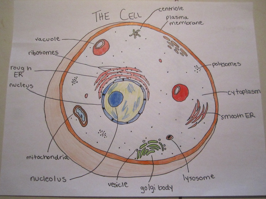
The Organelle's of the Cell
Nucleus:
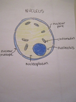
-The nucleus controls all cell activity.
-It also stores genetic information, and tells the cell which proteins to make.
-This process is called protein synthesis
-The nucleus has 5 parts: the nucleolus, chromatin/chromosomes, nuclear envelope, nucleoplasm, and the nuclear pores.
1. Nucleolus: The nucleolus makes ribosomes, RNA and has no membrane.
2. Chromatin/ Chromosomes: tightly coiled DNA, X shaped, wrapped around histone proteins, store genetic information.
3. Nuclear envelope: double layered membrane, covered with pores, and controls what enters nucleus.
4. Nuclear pores: holes through which RNA and proteins enter the nucleus. These are the only things allowed in to the nucleus.
5. Nucleoplasm: The liquid that fills the nucleus. It holds the chromatin, and the nucleolus.
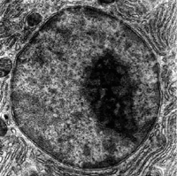
-Below is an electronmicrograph of the nucleus.
Mitochondria:
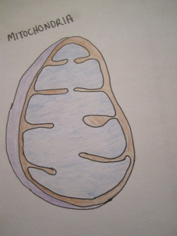
-The mitochondria is the furnace or energy maker of the cell.
-The process by which it makes energy is callled CELLULAR RESPIRATION.
The formula for cellular respiration is:
-sugar/glucose ----------------------> ATP Energy
C6H12O6 + O2 ---------------------> CO2 + H2O + ATP Energy
There is a high concentration of mitochondrias in the muscle tissue of the body, where a great deal of energy is necessary.
***Distinguishable features: The mitochondria has a swimming pool appearance. The large amounts of surface area allow for more area for cellular respiration to take place.
-Below is an electron micrograph of a mitochondria.
The Rough Endoplasmic Reticulum:
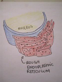
-The Rough ER is a transport system.
-It is covered with "dots" , which are actually ribosomes.
-Ribosomes make proteins, which the Rough ER transports OUTSIDE the cell.
-The rough ER is situated beside the nucleus.
-There is a high concentration of Rough ERs in the pancreas where the protein insulin is made.
***Distinguishable features: The Rough ER is covered with "dots", has a squished pancake appearance, and is beside the nucleus in the cell.
-Below is an electron micrograph of a Rough ER
The Smooth Endoplasmic Reticulum:
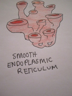
-The Smooth ER is another transport system.
-It transports lipids, hormones and steroids around the cell .
-It has NO ribosomes, but instead makes lipids and steroids.
-It also detoxifies the cell.
-There is an abundance of Smooth ERs in the female reproduction organs, because Smooth ERs create the necessary hormones.
***Distinguishable features: looks like rough ER, but without the ":dots".
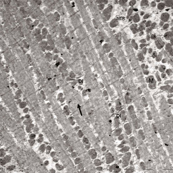
-Below is an electron micrograph of a Smooth ER
-It transports lipids, hormones and steroids around the cell .
-It has NO ribosomes, but instead makes lipids and steroids.
-It also detoxifies the cell.
-There is an abundance of Smooth ERs in the female reproduction organs, because Smooth ERs create the necessary hormones.
***Distinguishable features: looks like rough ER, but without the ":dots".
-Below is an electron micrograph of a Smooth ER
Ribosomes:
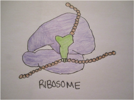
-Ribosomes make proteins for OUTSIDE the cell.
-They are situated on the rough ER, and make sure the amino acids are in the correct order.
-They read RNA, and then create the protein which the RNA codes for.
-As ribosomes make proteins, there are many ribosomes in the stomach where proteins are needed for digestion.
***Distinguishable features: Ribosomes are the "dots" on the Rough ER.
-Below is an electron micrograph of ribosomes.
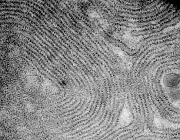
Polysome:
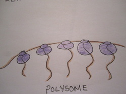
-A Polysome is a chain of ribosomes.
-They are seperate from the the Rough ER.
-They make a lot of the same type of protein for inside the cell.
-They appear as large clumps of dots in the cytoplasm.
-Polysomes are found in abundance in the digestive system.
**Distinguishable features: Polysomes are groups of "dots" NOT on the Rough ER, but in the cytoplasm.
-Below is an electron micrograph of polysomes.
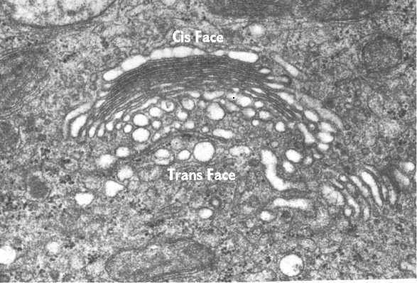
Golgi Body:
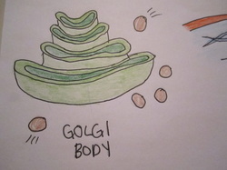
-The golgi body modifies proteins, hormones, and steroids.
-It also stores and packages them before they leave the cell.
-These are packaged in vesicles, and sent to where they are needed.
-There is a high concentration of golgi bodies in the stomach of the human body. Many proteins are needed to aid in the digestion process.
***Distinguishable features: The Golgi Body looks similar to the Smooth ER except it is slightly smaller.
-Below is an electronmicrograph of the golgi body.
Cytoskeleton:
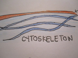
-The cytoskeleton gives shape to the cell, holds organelles and acts as a transport system for them.
-Think of a large subway station, running all around the cell.
-Cytoskeleton has 2 parts: microtubules and microfilaments.
-Microtubules: Hollow tubes, which work as a sort of scaffolding and give the cell its shape.
-Microfilaments: Very fine threads, mainly maid of the protein actin, which help with the movement of organelles within the cell.
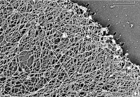
***Distinguishable features: Looks similar to a spider net, tangled, thin lines under the plasma membrane.
-Below is an electron micrograph of cytoskeleton.
-Think of a large subway station, running all around the cell.
-Cytoskeleton has 2 parts: microtubules and microfilaments.
-Microtubules: Hollow tubes, which work as a sort of scaffolding and give the cell its shape.
-Microfilaments: Very fine threads, mainly maid of the protein actin, which help with the movement of organelles within the cell.
***Distinguishable features: Looks similar to a spider net, tangled, thin lines under the plasma membrane.
-Below is an electron micrograph of cytoskeleton.
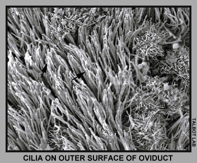
Cilia:

-Cilia are hair like projections.
-Cilia are shorter than flagella, are found in other cells besides the sperm cell.
-Their main function is to help in the movement of the cell and to clean.
-For example, there is a high concentration of cilia in your nostrils. They capture the dust and debris in the air, supplying your lungs with clean air.
-Below is an electronmicrograph of cilia.
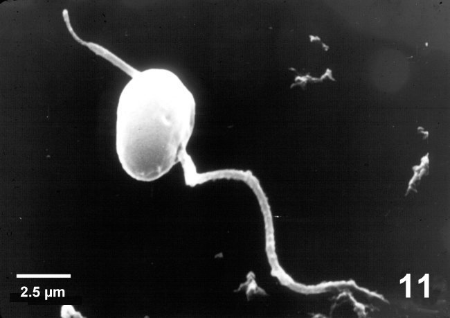
Flagella:
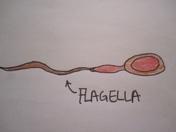
-The Flagella is a hair like projection, like the cillia, however it is longer.
-Their main function is to help propel the sperm cells forward. The long tale-like hair whips back and forth pushing the sperm cell forward.
-In the human body flagella are found only on the sperm cell, so there is a high concentration in semen.
-Below is an electron micrograph of flagella.
Centriole:
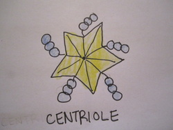
-Centrioles pull apart chromosomes during mitosis.
-They only exist in animal cells, and grow spindle fibres, which attach to the chromosomes and pull them apart during cell seperation.
***Distinguishable features: Centrioles are star shaped, their sides are made up of three tubes.
-Below is an electron micrograph of a centrioles
.
Cytoplasm:
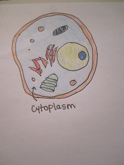
-Cytoplasm is watery jelly which fills the cell and gives it shape.
-It is filled with salts, proteins, and other things.
-Cytoplasm provides water for reactions within the cell.
-Below is an electronmicrograph of cytoplasm.
Organelle Relationships:
Cell organelles must work together to do various jobs. In order to understand the relationships between cell organelles, you must thoroughly understand the functions of the many cell organelles.
For example:
The nucleus, rough endoplasmic reticulum, vesicles, Golgi body, and cell membrane all work together. The nucleus gives the order to the cell to make a certain protein. The ribosomes on the Rough ER create the desired protein, which is then transported in a vesicle to the Golgi body. At the Golgi body the protein is stored until it is needed. At this time the golgi body may alter the order of the amino acids, but then the protein will packaged into another vesicle which will carry it to the cell membrane. At the cell membrane the vesicle will fuse with the cell membrane and expel its contents outside of the cell. This process is called exocytosis.
Questions:
1. What is the purpose of protein carriers on the plasma membrane?
2. What are the five parts of the nucleus?
3. What is the difference between polysomes and ribosomes?
4. What is the difference between the Rough ER and the Smooth ER? Include details about the appearance, location, and function.
5. What is the purpose of centrioles? What is notable about centriole's appearance?
6. Describe the relationships between the Rough ER, vesicles, and the cell memrane.
For additional help, please visit this website.
http://birkeland.weebly.com/screencasts.html